Diagnostic imaging lets doctors look inside your body for clues about a medical condition. A variety of machines and techniques can create pictures of the structures and activities inside your body. The type of imaging your doctor uses depends on your symptoms and the part of your body being examined.
Many imaging tests are painless and easy. Some require you to stay still for a long time inside a machine. This can be uncomfortable. Certain tests involve exposure to a small amount of radiation.
Below is a list of the most common diagnostic imaging procedures.
X-rays
What are X-rays?
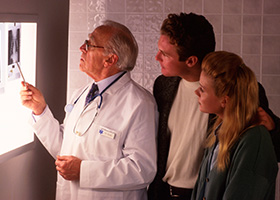 X-rays use invisible electromagnetic energy beams to produce images of internal tissues, bones, and organs on film or digital media. Standard X-rays are performed for many reasons, including diagnosing tumors or bone injuries. X-rays are made by using external radiation to produce images of the body, its organs, and other internal structures for diagnostic purposes. X-rays pass through body structures onto specially-treated plates (similar to camera film) or digital media and a “negative” type picture is made (the more solid a structure is, the whiter it appears on the film).
When the body undergoes X-rays, different parts of the body allow varying amounts of the X-ray beams to pass through. The soft tissues in the body (such as blood, skin, fat, and muscle) allow most of the X-ray to pass through and appear dark gray on the film or digital media. A bone or a tumor, which is more dense than soft tissue, allows few of the X-rays to pass through and appears white on the X-ray. When a break in a bone has occurred, the X-ray beam passes through the broken area and appears as a dark line in the white bone.
X-ray technology is used in other types of diagnostic procedures, such as arteriograms, computed tomography (CT) scans, and fluoroscopy.
Radiation during pregnancy may lead to birth defects. Always tell your radiologist or doctor if you suspect you may be pregnant.
How are X-rays performed?
X-rays can be performed on an outpatient basis, or as part of inpatient care.
Although each facility may have specific protocols in place, generally, an X-ray procedure follows this process:
- The patient will be asked to remove any clothing or jewelry which might interfere with the exposure of the body area to be examined. The patient will be given a gown to wear if clothing must be removed.
- The patient is positioned on an X-ray table that carefully positions the part of the body that is to be X-rayed–between the X-ray machine and a cassette containing the X-ray film or specialized image plate. Some examinations may be performed with the patient in a sitting or standing position.
- Body parts not being imaged may be covered with a lead apron (shield) to avoid exposure to the X-rays.
- The X-ray beam will be aimed at the area to be imaged.
- The patient must be very still or the image will be blurred.
- The technologist will step behind a protective window and the image is taken.
- Depending on the body part under study, various X-rays may be taken at different angles, such as the front and side view during a chest X-ray.
Computerized tomography (CT)
What is a CT or CAT scan?
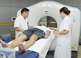
A CT or CAT scan is a diagnostic imaging procedure that uses a combination of X-rays and computer technology to produce horizontal, or axial, images (often called slices) of the body. A CT scan shows detailed images of any part of the body, including the bones, muscles, fat, organs, and blood vessels. CT scans are more detailed than standard X-rays.
In standard X-rays, a beam of energy is aimed at the body part being studied. A plate behind the body part captures the variations of the energy beam after it passes through skin, bone, muscle, and other tissue. While much information can be obtained from a regular X-ray, a lot of detail about internal organs and other structures is not available.
In computed tomography, the X-ray beam moves in a circle around the body. This allows many different views of the same organ or structure, and provides much greater detail. The X-ray information is sent to a computer that interprets the X-ray data and displays it in two-dimensional form on a monitor. Newer technology and computer software makes three-dimensional (3-D) images possible.
CT scans may be done with or without contrast. “Contrast” refers to a substance taken by mouth or injected into an intravenous (IV) line that causes the particular organ or tissue under study to be seen more clearly. Contrast examinations may require you to fast for a certain period of time before the procedure. Your doctor will notify you of this prior to the procedure.
CT scans may be performed to help diagnose tumors, investigate internal bleeding, or check for other internal injuries or damage.
You may want to ask your doctor about the amount of radiation used during the CT procedure and the risks related to your particular situation. It is a good idea to keep a record of your past history of radiation exposure, such as previous CT scans and other types of X-rays, so that you can inform your doctor. Risks associated with radiation exposure may be related to the cumulative number of X-ray examinations and/or treatments over a long period of time. If you are pregnant or suspect that you may be pregnant, you should notify your doctor.
Advances in computed tomography technology include the following:
- High-resolution computed tomography. This type of CT scan uses very thin slices (less than one-tenth of an inch), which are effective in providing greater detail in certain conditions such as lung disease.
- Helical or spiral computed tomography. During this type of CT scan, both the patient and the X-ray beam move continuously, with the X-ray beam circling the patient. The images are obtained much more quickly than with standard CT scans. The resulting images have greater resolution and contrast, thus providing more detailed information. Multidetector row helical CT scanners may be used to obtain information about calcium build-up inside the coronary arteries of the heart.
- Ultrafast computed tomography (also called electron beam computed tomography). This type of CT scan produces images very rapidly, thus creating a type of “movie” of moving parts of the body, such as the chambers and valves of the heart. This scan may also be used to obtain information about calcium build-up inside the coronary arteries of the heart, but the helical scanners are much more common.
- Computed tomographic angiography (CTA). Angiography (or arteriography) is an X-ray image of the blood vessels. A CT angiogram uses CT technology rather than standard X-rays or fluoroscopy to obtain images of blood vessels, for example, the coronary arteries of the heart.
- Combined computed tomography and positron emission tomography (PET/CT). The combination of computed tomography and positron emission tomography technologies into a single machine is referred to as PET/CT. PET/CT combines the ability of CT to provide detailed anatomy with the ability of PET to show cell function and metabolism to offer greater accuracy in the diagnosis and treatment of certain types of diseases, particularly cancer. PET/CT may also be used to evaluate epilepsy, Alzheimer’s disease, and coronary artery disease.
Studies show that 85 percent of the population will not experience an adverse reaction from iodinated contrast; however, you will need to let your doctor know if you have ever had a reaction to any contrast dye, and/or any kidney problems. A reported seafood allergy is not considered to be a contraindication for iodinated contrast. If you have any medical conditions or recent illnesses, inform your doctor. The effects of kidney disease and contrast agents have attracted increased attention over the last decade, as patients with kidney disease are more prone to kidney damage after contrast exposure. If you are pregnant or think you may be pregnant, you should notify your health care provider. If you are claustrophobic or tend to become anxious easily, tell your doctor ahead of time, as he or she may prescribe a mild sedative for you before the procedure to make you more comfortable. It will be necessary for you to remain still and quiet during the procedure, which may last 10 to 20 minutes.
How is a CT or CAT scan performed?
CT scans can be performed on an outpatient basis, unless they are part of a patient’s inpatient care. Although each facility may have specific protocols in place, generally, CT scans follow this process:
- When the patient arrives for the CT scan, he or she will be asked to remove any clothing, jewelry, or other objects that may interfere with the scan.
- If the patient will be having a procedure done with contrast, an intravenous (IV) line will be started in the hand or arm for injection of the contrast medication. For oral contrast, the patient will be given the contrast material to swallow.
- The patient will lie on a scan table that slides into a large, circular opening of the scanning machine.
- The CT staff will be in another room where the scanner controls are located. However, the patient will be in constant sight of the staff through a window. Speakers inside the scanner will enable the staff to communicate with and hear the patient. The patient may have a call bell so that he or she can let the staff know if he or she has any problems during the procedure.
- As the scanner rotates around the patient, X-rays will pass through the body for short amounts of time. The motion is hidden inside the gantry, the doughnut-shaped part of the machine. The patient may hear buzzing, whirring, and clicking as the X-ray tube rotates.
- The X-rays absorbed by the body’s tissues will be detected by the scanner and transmitted to the computer.
- The computer will transform the information into an image to be interpreted by the radiologist.
- It is very important that the patient remain very still during the procedure. You may be asked to hold your breath at various times during the procedure.
- The technologist will be watching the patient at all times and will be in constant communication.
- The patient may be asked to wait for a short period of time while the radiologist examines the scans to make sure they are clear. If the scans are not clear enough to obtain adequate information, the patient may need to have additional scans performed.
Positron Emission Tomography (PET)
What is PET?
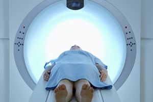 Positron emission tomography (PET) is a specialized radiology procedure used to examine various body tissues to identify certain conditions. PET may also be used to follow the progress of the treatment of certain conditions. While PET is most commonly used in the fields of neurology, oncology, and cardiology, applications in other fields are currently being studied.
PET is a type of nuclear medicine procedure. This means that a tiny amount of a radioactive substance, called a radionuclide (radiopharmaceutical or radioactive tracer), is used during the procedure to assist in the examination of the tissue under study. Specifically, PET studies evaluate the metabolism of a particular organ or tissue, so that information about the physiology (functionality) and anatomy (structure) of the organ or tissue is evaluated, as well as its biochemical properties. Thus, PET may detect biochemical changes in an organ or tissue that can identify the onset of a disease process before anatomical changes related to the disease can be seen with other imaging processes, such as computed tomography (CT) or magnetic resonance imaging (MRI).
PET is most often used by oncologists (doctors specializing in cancer treatment), neurologists and neurosurgeons (doctors specializing in treatment and surgery of the brain and nervous system), and cardiologists (doctors specializing in the treatment of the heart). However, as advances in PET technologies continue, this procedure is beginning to be used more widely in other areas.
PET is also being used in conjunction with other diagnostic tests such as computed tomography (CT) to provide more definitive information about malignant (cancerous) tumors and other lesions. The combination of PET and CT shows particular promise in the diagnosis and treatment of many types of cancer.
Until recently, PET procedures were performed in dedicated PET centers. The equipment used in these centers is quite expensive. However, a new technology called gamma camera systems (devices used to scan patients who have been injected with small amounts of radionuclides and currently in use with other nuclear medicine procedures) is now being adapted for use in PET scan procedures. The gamma camera system can complete a scan more quickly, and at less cost, than a traditional PET scan.
How does PET work?
PET works by using a scanning device (a machine with a large hole at its center) to detect positrons (subatomic particles) emitted by a radionuclide in the organ or tissue being examined.
The radionuclides used in PET scans are made by attaching a radioactive atom to chemical substances that are used naturally by the particular organ or tissue during its metabolic process. For example, in PET scans of the brain, a radioactive atom is applied to glucose (blood sugar) to create a radionuclide called fluorodeoxyglucose (FDG), because the brain uses glucose for its metabolism. FDG is widely used in PET scanning.
Other substances may be used for PET scanning, depending on the purpose of the scan. If blood flow and perfusion of an organ or tissue is of interest, the radionuclide may be a type of radioactive oxygen, carbon, nitrogen, or gallium.
The radionuclide is administered into a vein through an intravenous (IV) line. Next, the PET scanner slowly moves over the part of the body being examined. Positrons are emitted by the breakdown of the radionuclide. Gamma rays are created during the emission of positrons, and the scanner then detects the gamma rays. A computer analyzes the gamma rays and uses the information to create an image map of the organ or tissue being studied. The amount of the radionuclide collected in the tissue affects how brightly the tissue appears on the image, and indicates the level of organ or tissue function.
Other related procedures that may be performed include computed tomography (CT scan) and magnetic resonance imaging (MRI). Please see these procedures for additional information.
Reasons for the procedure
In general, PET scans may be used to evaluate organs and/or tissues for the presence of disease or other conditions. PET may also be used to evaluate the function of organs such as the heart or brain. Another use of PET scans is in the evaluation of the treatment of cancer.
More specific reasons for PET scans include, but are not limited to, the following:
- To diagnose dementias such as Alzheimer’s disease, as well as other neurological conditions such as Parkinson’s disease (a progressive disease of the nervous system in which a fine tremor, muscle weakness, and a peculiar type of gait are seen), Huntington’s disease (a hereditary disease of the nervous system which causes increasing dementia, bizarre involuntary movements, and abnormal posture), epilepsy (a brain disorder involving recurrent seizures), and cerebrovascular accident (stroke)
- To locate the specific surgical site prior to surgical procedures of the brain
- To evaluate the brain after trauma to detect hematoma (blood clot), bleeding, and/or perfusion (blood and oxygen flow) of the brain tissue
- To detect the spread of cancer to other parts of the body from the original cancer site
- To evaluate the effectiveness of cancer treatment
- To evaluate the perfusion to the myocardium (heart muscle) as an aid in determining the usefulness of a therapeutic procedure to improve blood flow to the myocardium
- To further identify lung lesions or masses detected on chest X-ray and/or chest CT
- To assist in the management and treatment of lung cancer by staging lesions and following the progress of lesions after treatment
- To detect recurrence of tumors earlier than with other diagnostic modalities
There may be other reasons for your doctor to recommend a PET scan.
Risks of the procedure
The amount of the radionuclide injected into your vein for the procedure is small enough that there is no need for precautions against radioactive exposure. The injection of the radionuclide may cause some slight discomfort. Allergic reactions to the radionuclide are rare, but may occur.
For some patients, having to lie still on the scanning table for the length of the procedure may cause some discomfort or pain.
Patients who are allergic to or sensitive to medications, contrast dyes, iodine, or latex should notify their doctor.
If you are pregnant or suspect that you may be pregnant, you should notify your health care provider due to the risk of injury to the fetus from a PET scan. If you are lactating, or breastfeeding, you should notify your health care provider due to the risk of contaminating breast milk with the radionuclide.
There may be other risks depending on your specific medical condition. Be sure to discuss any concerns with your doctor prior to the procedure.
Certain factors or conditions may interfere with the accuracy of a PET scan. These factors include, but are not limited to, the following:
- High blood glucose levels in diabetics
- Caffeine, alcohol, or tobacco consumed within 24 hours of the procedure
- Medications, such as insulin, tranquilizers, and sedatives
Notify your doctor if any of the above situations may apply to you.
Before the procedure
- Your doctor will explain the procedure and offer you the opportunity to ask any questions that you might have about the procedure.
- You may be asked to sign a consent form that gives your permission to do the procedure. Read the form carefully and ask questions if something is not clear.
- Notify the radiologist or technologist if you are allergic to latex and/or sensitive to medications, contrast dyes, or iodine.
- Fasting for a certain period of time prior to the procedure is generally required. Your doctor will give you special instructions ahead of time as to the number of hours you are to withhold food and drink. Your doctor will also inform you as to the use of medications prior to the PET scan.
- Notify your doctor if you are pregnant or suspect you may be pregnant.
- Notify your doctor of all medications (prescribed and over-the-counter) and herbal supplements that you are taking.
- You should not consume any caffeine or alcohol, or use tobacco, for at least 24 hours prior to the procedure.
- If you are a diabetic who uses insulin, you may be instructed to take your preprocedure insulin dose with a meal several hours prior to the procedure. Your doctor will give you specific instructions based on your individual situation. Also, a fasting blood sugar test may be obtained prior to the procedure. If your blood sugar is elevated, you may be given insulin to lower the blood sugar.
- Based on your medical condition, your doctor may request other specific preparation.
During the procedure
PET scans may be performed on an outpatient basis or as part of your stay in a hospital. Procedures may vary depending on your condition and your doctor’s practices.
Generally, a PET scan follows this process:
- You will be asked to remove any clothing, jewelry, or other objects that may interfere with the scan.
- If you are asked to remove clothing, you will be given a gown to wear.
- You will be asked to empty your bladder prior to the start of the procedure.
- One or two intravenous (IV) lines will be started in the hand or arm for injection of the radionuclide.
- Certain types of scans of the abdomen or pelvis may require that a urinary catheter be inserted into the bladder to drain urine during the procedure.
- In some cases, an initial scan may be performed prior to the injection of the radionuclide, depending on the type of study being done. You will be positioned on a padded table inside the scanner.
- The radionuclide will be injected into your vein. The radionuclide will be allowed to concentrate in the organ or tissue for about 30 to 60 minutes. You will remain in the facility during this time. You will not be hazardous to other people, as the radionuclide emits less radiation than a standard X-ray.
- After the radionuclide has been absorbed for the appropriate length of time, the scan will begin. The scanner will move slowly over the body part being studied.
- When the scan has been completed, the IV line will be removed. If a urinary catheter has been inserted, it will be removed.
While the PET scan itself causes no pain, having to lie still for the length of the procedure might cause some discomfort or pain, particularly in the case of a recent injury or invasive procedure, such as surgery. The technologist will use all possible comfort measures and complete the procedure as quickly as possible to minimize any discomfort or pain.
After the procedure
You should move slowly when getting up from the scanner table to avoid any dizziness or lightheadedness from lying flat for the length of the procedure.
You will be instructed to drink plenty of fluids and empty your bladder frequently for 24 to 48 hours after the test to help flush the remaining radionuclide from your body.
The IV site will be checked for any signs of redness or swelling. If you notice any pain, redness, and/or swelling at the IV site after you return home following your procedure, you should notify your doctor as this may indicate an infection or other type of reaction.
Your doctor may give you additional or alternate instructions after the procedure, depending on your particular situation.
Fluoroscopy
Procedure overview
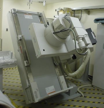 Fluoroscopy is a study of moving body structures–similar to an X-ray “movie.” A continuous X-ray beam is passed through the body part being examined. The beam is transmitted to a TV-like monitor so that the body part and its motion can be seen in detail. Fluoroscopy, as an imaging tool, enables physicians to look at many body systems, including the skeletal, digestive, urinary, respiratory, and reproductive systems. Fluoroscopy may be performed to evaluate specific areas of the body, including the bones, muscles, and joints, as well as solid organs, such as the heart, lung, or kidneys.Other related procedures that may be used to diagnose problems of the bones, muscles, or joints include X-rays, myelography (myelogram), computed tomography (CT scan), magnetic resonance imaging (MRI), and arthrography. Please see these procedures for additional information.
Photo by: Normaler Schluckakt
Reasons for the procedure
Fluoroscopy is used in many types of examinations and procedures, such as barium X-rays, cardiac catheterization, arthrography (visualization of a joint or joints), lumbar puncture, placement of intravenous (IV) catheters (hollow tubes inserted into veins or arteries), intravenous pyelogram, hysterosalpingogram, and biopsies.
Fluoroscopy may be used alone as a diagnostic procedure, or may be used in conjunction with other diagnostic or therapeutic media or procedures.
In barium X-rays, fluoroscopy used alone allows the doctor to see the movement of the intestines as the barium moves through them and allows the doctor to position the patient for spot imaging. In cardiac catheterization, fluoroscopy is used as an adjunct to enable the doctor to see the flow of blood through the coronary arteries in order to evaluate the presence of arterial blockages. For intravenous catheter insertion, fluoroscopy assists the doctor in guiding the catheter into a specific location inside the body.
Other uses of fluoroscopy include, but are not limited to, the following:
- Locating foreign bodies
- Image-guided anesthetic injections into joints or the spine
- Percutaneous vertebroplasty. A minimally invasive procedure used to treat compression fractures of the vertebrae of the spine
There may be other reasons for your doctor to recommend fluoroscopy.
Risks of the procedure
You may want to ask your doctor about the amount of radiation used during the procedure and the risks related to your particular situation. It is a good idea to keep a record of your past history of radiation exposure, such as previous scans and other types of X-rays, so that you can inform your doctor. Risks associated with radiation exposure may be related to the cumulative number of X-ray examinations and/or treatments over a long period of time.
If you are pregnant or suspect that you may be pregnant, you should notify your doctor. Radiation exposure during pregnancy may lead to birth defects.
If contrast dye is used, there is a risk for allergic reaction to the dye. Patients who are allergic to or sensitive to medications, contrast media, iodine, or latex should notify their doctor. Also, patients with kidney failure or other kidney problems should notify their doctor.
There may be other risks depending on your specific medical condition. Be sure to discuss any concerns with your doctor prior to the procedure.
Certain factors or conditions may interfere with the accuracy of a fluoroscopy procedure. A recent barium X-ray procedure may interfere with exposure of the abdominal or lower back area.
Before the procedure
- Your doctor will explain the procedure to you and offer you the opportunity to ask any questions that you might have about the procedure.
- You may be asked to sign a consent form that gives your permission to do the procedure. Read the form carefully and ask questions if something is not clear.
- The specific type of procedure or examination being done will determine whether any preparation prior to the procedure is required. Your doctor will notify you of any preprocedure instructions.
- Notify your doctor if you have ever had a reaction to any contrast dye, or if you are allergic to iodine.
- If you are pregnant or suspect that you may be pregnant, you should notify your doctor.
- Based on your medical condition, your doctor may request other specific preparation.
During the procedure
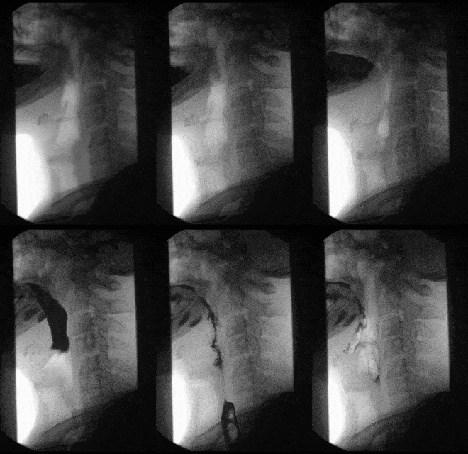 Fluoroscopy may be performed on an outpatient basis or as part of your stay in a hospital. Procedures may vary depending on your condition and your doctor’s practices.
Generally, fluoroscopy follows this process:
- You will be asked to remove any clothing or jewelry that may interfere with the exposure of the body area to be examined.
- If you are asked to remove clothing, you will be given a gown to wear.
- A contrast substance may be given, depending on the type of procedure that is being performed, via swallowing, enema, or an intravenous (IV) line in your hand or arm.
- You will be positioned on the X-ray table. Depending on the type of procedure, you may be asked to assume different positions, move a specific body part, or hold your breath at intervals while the fluoroscopy is being performed.
- For procedures that require catheter insertion, such as cardiac catheterization or catheter placement into a joint or other body part, an additional line insertion site may be used in the groin, elbow, or other site.
- A special X-ray machine will be used to produce the fluoroscopic images of the body structure being examined or treated.
- A dye or contrast substance may be injected into the IV line in order to better visualize the organs or structures being studied.
- In the case of arthrography (visualization of a joint), any fluid in the joint may be aspirated (withdrawn with a needle) prior to the injection of the contrast substance. After the contrast is injected, you may be asked to move the joint for a few minutes in order to evenly distribute the contrast substance throughout the joint.
- The type of procedure being performed and the body part being examined and/or treated will determine the length of the procedure.
- After the procedure has been completed, the IV line will be removed.
While fluoroscopy itself is not painful, the particular procedure being performed may be painful, such as the injection into a joint or accessing of an artery or vein for angiography. In these cases, the radiologist will take all comfort measures possible, which could include local anesthesia, conscious sedation, or general anesthesia, depending on the particular procedure.
After the procedure
The type of care required after the procedure will depend on the type of fluoroscopy that is performed. Certain procedures, such as cardiac catheterization, will likely require a recovery period of several hours with immobilization of the leg or arm where the cardiac catheter was inserted. Other procedures may require less time for recovery.
If you notice any pain, redness, and/or swelling at the IV site after you return home following your procedure, you should notify your doctor as this could indicate an infection or other type of reaction.
Your doctor will give more specific instructions related to your care after the examination or procedure.
Magnetic Resonance Imaging (MRI)
How does an MRI scan work?
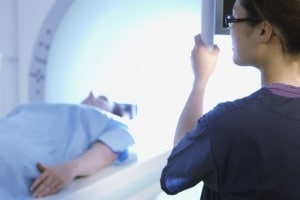 The MRI machine is a large, cylindrical (tube-shaped) machine that creates a strong magnetic field around the patient and sends pulses of radio waves from a scanner. The strong magnetic field causes the hydrogen atoms in your body to align along the same axis. The radio waves knock the nuclei of the atoms in your body out of this aligned position. As the nuclei realign back into proper position, they send out radio signals. These signals are received by a computer that analyzes and converts them into an image of the part of the body being examined. This image appears on a viewing monitor. Cross-sectional views can be obtained to reveal further details. Some MRI machines look like narrow tunnels, while others are more open.
Magnetic resonance imaging (MRI) may be used instead of computed tomography (CT) in situations where organs or soft tissue are being studied, because MRI is better at telling the difference between different types of soft tissues, as well as the difference between normal and abnormal soft tissues..
Because ionizing radiation is not used, there is no risk of exposure to ionizing radiation during an MRI procedure.
Due to the use of the strong magnet, MRI cannot be performed on most patients with implanted pacemakers, older intracranial aneurysm clips, cochlear implants, certain prosthetic devices, implanted drug infusion pumps, neurostimulators, bone-growth stimulators, certain intrauterine contraceptive devices, or any other type of iron-based metal implants. MRI is also not recommended for people who have internal metallic objects such as bullets or shrapnel, as well as most surgical clips, pins, plates, screws, metal sutures, or wire mesh in their bodies. Dyes used in tattoos may contain iron and potentially could heat up during an MRI, but this is a rare occurrence.
Newer uses and indications for MRI have contributed to the development of additional magnetic resonance technology. Magnetic resonance angiography (MRA) is a procedure used to evaluate blood flow through arteries in a noninvasive (the skin is not pierced) manner. MRA can also be used to detect aneurysms within the brain and vascular malformations (abnormalities of blood vessels within the brain, spinal cord, or other parts of the body).
Magnetic resonance spectroscopy (MRS) is another noninvasive procedure used to assess chemical abnormalities in body tissues such as the brain. MRS may be used to assess disorders, such as HIV infection of the brain, stroke, head injury, coma, Alzheimer’s disease, tumors, and multiple sclerosis.
Functional magnetic resonance imaging of the brain (fMRI) is used to determine the specific location within the brain where a certain function, such as speech or memory, occurs. The general areas of the brain in which such functions occur are known, but the exact location may vary from person to person. During functional resonance imaging of the brain, you will be asked to perform a specific task, such as recite the Pledge of Allegiance, while the scan is being done. By pinpointing the exact location of the functional center in the brain, doctors can plan surgery or other treatments for a particular disorder of the brain.
Another advance in MRI technology is the “open” MRI. Standard MRI units have a closed cylinder-shaped tunnel into which the patient is placed for the procedure. Open MRI units do not completely surround the patient, and some units may be open on all sides. Open MRI units are particularly useful for procedures involving:
- Children. Parents or other caregivers may stay with a child during the procedure to provide comfort and security.
- Claustrophobia. Before the development of open MRI units, persons with severe claustrophobia often required a sedative medication prior to the procedure.
- Very large or obese persons. Almost anyone can be accommodated in most open MRI units.
How is an MRI performed?
An MRI may be performed on an outpatient basis, or as part of inpatient care. Although each facility may have specific protocols in place, generally, an MRI procedure follows this process:
- Because of the strong magnetic field, the patient must remove all jewelry and metal objects, such as hairpins or barrettes, hearing aids, eyeglasses, and dental pieces.
- If a contrast medication and/or sedative are to be given by an intravenous line (IV), an IV line will be started in the hand or arm. If the contrast is to be taken by mouth, the patient will be given the contrast to swallow.
- The patient will lie on a table that slides into a tunnel in the scanner.
- The MRI staff will be in another room where the scanner controls are located. However, the patient will be in constant sight of the staff through a window. Speakers inside the scanner will enable the staff to communicate with and hear the patient. The patient will have a call bell so that he or she can let the staff know if he or she has any problems during the procedure.
- During the scanning process, a clicking noise will sound as the magnetic field is created and pulses of radio waves are sent from the scanner. The patient may be given headphones to wear to help block out the noises from the MRI scanner and hear any messages or instructions from the technologist.
- It is important that the patient remain very still during the examination.
- At intervals, the patient may be instructed to hold his or her breath, or to not breathe, for a few seconds, depending on the body part being examined. The patient will then be told when he or she can breathe. The patient should not have to hold his or her breath for longer than a few seconds, so this should not be uncomfortable.
- The technologist will be watching the patient at all times and will be in constant communication.
Ultrasound
What is an ultrasound?
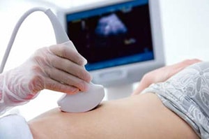
An ultrasound procedure is a noninvasive (the skin is not pierced) diagnostic procedure used to assess soft tissue structures such as muscles, blood vessels, and organs. Ultrasound uses a transducer that sends out ultrasonic sound waves at a frequency too high to be heard. When the transducer is placed at certain locations and angles, the ultrasonic sound waves move through the skin and other body tissues to the organs and structures within. The sound waves bounce off the organs like an echo and return to the transducer. The transducer picks up the reflected waves, which are then converted by a computer into an electronic picture of the organs or tissues under study.
Different types of body tissues affect the speed at which sound waves travel. Sound travels the fastest through bone tissue, and moves most slowly through air. The speed at which the sound waves are returned to the transducer, as well as how much of the sound wave returns, is translated by the transducer as different types of tissue. A clear conducting gel is placed between the transducer and the skin to allow for smooth movement of the transducer over the skin and to eliminate air between the skin and the transducer for the best sound conduction. By using an additional mode of ultrasound technology during an ultrasound procedure, blood flow can be assessed. An ultrasound transducer capable of assessing blood flow contains a Doppler probe.
The Doppler probe within the transducer evaluates the velocity and direction of blood flow in the vessel and makes the sound waves audible. Ultrasounds are used to view internal organs as they function (in “real time,” like a live TV broadcast), and to assess blood flow through various vessels. Ultrasound procedures are often used to examine many parts of the body such as the abdomen, breasts, female pelvis, prostate, scrotum, thyroid and parathyroid glands, and the vascular system. During pregnancy, ultrasounds are performed to evaluate the development of the fetus. Technological advancements in the field of ultrasound now include images that can be made in a three-dimensional view (3-D) and/or four dimensional (4-D) view. The added dimension of the 4-D is motion, so that it is a 3-D view with movement.
What are the different types of ultrasound procedures?
Different ultrasound techniques exist for different conditions. Examples of some of the more common types of ultrasound examinations include the following:
- Doppler ultrasound. Used to see structures inside the body, while evaluating blood flow at the same time. Doppler ultrasound can determine if there are any problems within the veins and arteries.
- Vascular ultrasound. Used to see the vascular system and its function, including detection of blood clots.
- Echocardiogram. Used to see the heart and its valves, and to evaluate the effectiveness of the heart’s pumping ability.
- Abdominal ultrasound. Used to detect any abnormalities of the abdominal organs (i.e., kidneys, liver, pancreas, gallbladder), such as gallstones or tumors.
- Renal ultrasound. Used to examine the kidneys and urinary tract.
- Obstetrical ultrasound. Used to monitor the development of the fetus.
- Pelvic ultrasound. Used to find the cause of pelvic pain, such as an ectopic pregnancy in women, or to detect tumors or masses.
- Breast ultrasound. Used to examine a mass in the breast tissue.
- Thyroid ultrasound. Used to see the thyroid and to detect any abnormalities.
- Scrotal ultrasound. Used to further investigate pain in the testicles.
- Prostate ultrasound. Used to examine any nodules felt during a physical examination.
- Musculoskeletal ultrasound. Used to examine any joint or muscle pain for conditions, such as a tear.
- Intraoperative ultrasound. Used to help the surgeon during a minimally invasive operation or biopsy.
- Interventional ultrasound. Used by an interventional radiologist to guide a minimally invasive procedure.
- Intravascular ultrasound (IVUS). Used to provide direct visualization and measurement of the inside of blood vessels.
- Endoscopic ultrasound. Used to obtain direct ultrasound examination of the inside of a body cavity or organ, using an ultrasound transducer inside an endoscope (a small, flexible tube with a light and a lens on the end).
How are ultrasounds performed?
An ultrasound procedure may be done on an outpatient basis, or as part of inpatient care. Although each facility may have specific protocols in place, generally, an ultrasound procedure follows this process:
- A gel-like substance will be smeared on the area of the body to undergo the ultrasound (the gel acts as a conductor).
- Using a transducer, a tool that sends ultrasound waves, the ultrasound will be sent through the patient’s body.
- The sound from the transducer will be reflected off structures inside the body, and the information from the sound waves will be analyzed by a computer.
- The computer will create an image of these structures on a television screen. The moving pictures can be recorded.
- There are no confirmed adverse biological effects on patients or instrument operators caused by exposures to ultrasound.


